


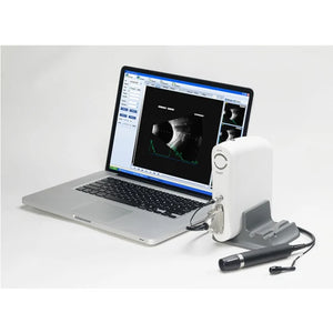
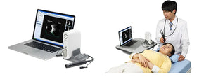

Functions of Ophthalmology A-B Scanner:
- Software image workstation
- B, B+B, B+A, A modes
- Video review for 100 images
- PDF report output
- Optional 20MHz B Probe: vitreous plus function
Technical Specifications:
| A scan | 1.Probe: 10MHz frequencies, with LED 2.Depth: 40mm 3.Precision: ±0.05mm 4.Eye mode: Phakic / Aphakic / Dense / Various IOL 5.Measurement: Anterior chamber depth, lens thickness, vitreous body length, total length and average 6.IOL Formula: SRK-II, SRK-T, BINKHORST, HOLLADAY, HOFFER-Q, HAIGIS, Stat. 7.Calculation: Average and standard deviation 8.Store: 10 Scanning results for each eye |
| B scan | 1.Probe: 10MHz/20MHz (optional), Magnetic driven, noiseless 2.Scanning Mode: Sector Scanning 3.Resolution: Lateral ≤0.3mm; Vertical≤0.2mm 4.Geometric Location Precision: Lateral≤10%; Vertical≤5% 5.Depth: 60mm 6.Enhance the part of vitreous body and retina 7.Gain of probe:30dB-105dB 8.Scanning Angle : 53° 9.Gray Scale: 256 10.False Color: Multi colors OTC 11.Measure Mode: distances, perimeter and area 12.Movies: 100 images movie review,AVI ZIP JPG format image output 13.Output: PDF format case report, connect to normal printer |
| Others | 1.Display Mode :B, B+B, B+A, A 2.Hint: preset keyword 3.Case Search: Multi-keywords 4.Working Platform: Windows XP, VISTA, WINDOWS7 5.User-defined report template |